

Insider Brief
- Researchers at the University of Göttingen have developed a method to map the three-dimensional wave function of a nanometer-scale organic molecule using a laboratory-scale soft X-ray source.
- The approach combines photoelectron spectroscopy with a redesigned computational algorithm to reconstruct molecular orbitals with sub-carbon-atom resolution from reduced measurement data.
- The researchers say the method could enable femtosecond-scale imaging of how molecular wave functions change in response to optical, electronic and chemical stimuli.
Press release – Electrons cannot be fixed to a single location. This is one of the central insights of quantum mechanics. The wave function takes the place of the location: a mathematical quantity that contains all the important information about the particle. An interdisciplinary research team from the University of Göttingen has now succeeded in mapping the wave function of a nanometer-sized organic molecule three-dimensionally – using an X-ray light source that fits on a laboratory table. The results were published in the journal Nature Communications.
“The wave function is a fundamental quantity in quantum mechanics, but cannot be measured directly“, explains Prof. Dr. Stefan Mathias, Head of the Research Group “Ultrafast Dynamics in Quantum Materials“ at the University of Göttingen. Of particular interest are the wave functions of electrons in molecules, the so-called molecular orbitals: they determine how a molecule absorbs light or undergoes chemical reactions.
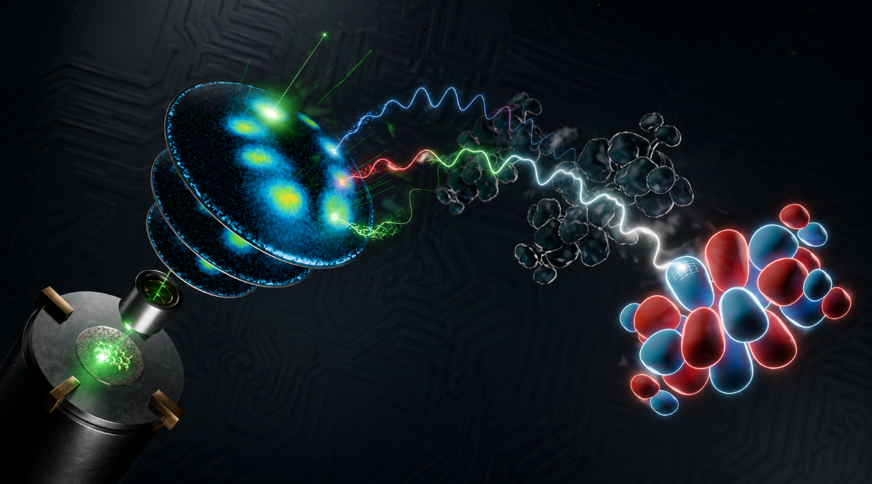
To make them visible, the team combined high-precision photoelectron spectroscopy –in which light releases electrons from the material whose momentum allows conclusions to be drawn about the wave function – with powerful computer algorithms. The result: a complete 3D image of the molecular orbital, with a resolution finer than the distance between two carbon atoms.Until now, such recordings were only possible at large accelerator facilities, so-called synchrotrons, and even there only with considerable time expenditure.
“We introduce two new concepts: Firstly, our completely redesigned algorithm delivers reliable 3D images from significantly less measurement data. Secondly, the experiment is based on a powerful laboratory-scale light source that generates ultrashort pulses of soft X-rays“, explains Dr. Matthijs Jansen, member of the research group „Ultrafast Dynamics in Quantum Materials“ and one of the leaders of the study. „It is the combination of both methods that makes these results so impressive.“ First author Dr. Wiebke Bennecke from the same research group adds: “This could make stroboscopic videography a reality: We would not only see the shape of wave functions, but also how they change – with a time resolution of femtoseconds, i.e. billiard parts of seconds. This is how we learn,how a molecule reacts to optical, electronic, or chemical stimuli, and find new ways to control these processes on an atomic scale.“
Original publication: Bennecke, W. et al. “Table-top three-dimensional photoemission orbital tomography with a femtosecond extreme ultraviolet light source.” Nature Communications (2026). DOI: 10.1038/s41467-026-74308-1